
Disclaimer: this guide does not replace consultation with your health care professional. Full disclaimer.
What is a biopsy used for?
A breast biopsy is a diagnostic test which involves the removal of a small sample of breast tissue from a suspicious area, which is then sent off to be examined in a lab by a pathologist. The main reason a breast biopsy is performed is to determine whether a lump is cancerous.
Biopsy methods
Breast biopsy methods include fine needle aspiration (FNA), core needle biopsy (CNB), vacuum assisted biopsy (VAB), surgical biopsy and skin punch biopsy. Based on prebiopsy imaging, the doctor (radiologist) makes an assessment of which biopsy approach will yield the highest likelihood of success, while considering your safety and comfort. The choice of imaging guidance depends upon the modality on which the lump (lesion) is best visualized and whether it is palpable (can be felt or touched). The doctor will consider your tolerance for positioning required for the various imaging techniques. (e.g., lying face down is usually need for stereotactic or MRI guidance). The requirements of your potential subsequent therapy are additional factors that are included in the biopsy planning.18
Fine Needle Aspiration (FNA)
FNA, may be done when the doctor can feel or touch a lump (it is palpable), or it can be done under imaging (e.g., ultrasound) that show an abnormal growth area. FNA is the least invasive of all the biopsies, and can provide rapid preliminary diagnosis of cancer, which may expedite planning for treatment. However, because FNA often has higher rates of false negative results and insufficient samples sizes collected, CNB following FNA is sometimes necessary.18 The sensitivity of FNA for cancer diagnosis is lower than that for CNB. In a systematic review/meta-analysis, FNA demonstrated a sensitivity of 74% (95% CI 72 to 77%) and specificity of 96% (95% CI 94 to 98%)23. However, the accuracy varies with proceduralist experience and training.24
Procedure:
FNA is done in a doctor’s office or as an outpatient in a hospital. You usually don’t need any special preparation for the test, and the procedure does not take too long. The doctor may use local anesthetic to numb the area. They will then insert a very thin needle (usually attached to a syringe) through the skin and into the area being examined. Your doctor will slowly draw cells, tissue or fluid through the needle into the syringe. You may feel some pressure or discomfort during the FNA, but it’s not usually painful. Sometimes multiple samples from different areas are required to make a diagnosis. New needles and syringes are used for each FNA.22
Side effects include discomfort or soreness, bleeding, infection, potential damage to nearby structures.22 As FNA is done with a very small-sized needle, it very rarely leaves a scar.

Core Needle Biopsy (CNB)
In a CNB procedure, your doctor will remove a small but long narrow piece (called a "core") that is slightly larger than one that would be used for an FNA. The tissue remains intact and gives the doctor additional information about the tissue surrounding the cancer cells, and therefore they can decide if further tests or treatments are needed. This is an advantage over FNA, which does not preserve the tissue structure.18
In a meta-analysis, CNB under ultrasound or stereotactic guidance demonstrated a sensitivity of 87% (95% CI 84 to 88%) and specificity of 98% (95% CI 96 to 99%).18,23A core biopsy is performed in a doctor’s office, clinic or hospital. In most cases, you don’t need any special preparation and the procedure usually takes about 15-30 minutes. The procedure is performed under local anesthesia to freeze the area.18,25
CNB is performed using guidance with either ultrasound, x-ray (i.e., stereotactic or tomosynthesis), or MRI without and with intravenous contrast. In most cases your doctor will use ultrasound imaging to help guide the procedure (if the target lesion can be well visualized). Stereotactic or tomosynthesis biopsy is performed for mammographic abnormalities without a clear ultrasound correlate. MRI-guided biopsy is available at some sites for lesions seen only on MRI.18
A small skin incision is made through which a core biopsy needle is introduced. The shortest path to the lesion is typically chosen. The doctor will usually take several samples of tissue during the biopsy.25 Sometimes, during the procedure your doctor will ‘tag’ the suspect area using a very small marker, so that if it is determined that surgery is required, the affected region will be easier for the surgeon to locate.18, 20
Side effects may include tenderness, bleeding or bruising and general soreness of the breast for a short time after the procedure. The potential for infection is also possible.25 As the needle used in a core needle biopsy is slightly larger compared to FNA, it may leave a small scar.

Vacuum-assisted Breast Biopsy (VAB)
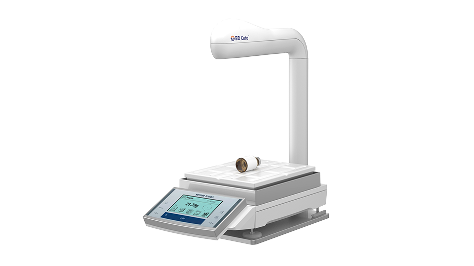
Your doctor may also recommend that you have a VAB, another minimally invasive core needle biopsy procedure. VAB is often used with stereotactic (mammogram-guided) or MRI imaging but can also be used with ultrasound. The use of VAB increases the volume of tissue that can be obtained quickly and may decrease the false negative rate (higher accuracy) for cancer detection.18,26,27,28,29,30 Reports of false negative rates low and have shown to range from 3.3% to lower than 0.45% , in several studies.16 If a second biopsy is required, due to inclusive first biopsy results using a FNA or CNB, a VAB will often be used because of its high accuracy. VAB can also be used instead of a surgical biopsy procedure, as it is quicker, less expensive and less painful for you.34,35,36
A VAB appointment generally takes on average less than 30 minutes . This can vary depending on where the biopsy is performed (i.e., hospital or radiology practice). During the procedure, a small ‘nick’ is made in your skin, about the size of half a grain of rice. The needle used in a VAB procedure is called a “probe”, which features a hollow needle connected to a vacuum pump or system. The probe is then inserted into your breast, guided with either ultrasound, stereotactic, or MRI. A gentle vacuum draws tissue inside the probe, while the needle rotates to takes multiple samples.18 VAB can efficiently collect several samples in a rotational fashion with only a single insertion, meaning the chances of obtaining an adequate tissue sample is greater.29
Just like with a CNB procedure, your doctor may also ‘tag’ the area with a very small marker for easier location in the future. Different shapes or types of tags may be used if more than one lesion is biopsied in order to differentiate the various sites. This become especially important in the event that surgery is required for only one of your lesions.18
Like FNA and CNB, side effects include minor bleeding, breast bruising and tenderness following the procedure.37 Scarring was rare among patients (0.3% of the 2874 patients had relevant scaring in one study.32 Although the size of a VAB needle is larger than a CNB needle, only a single insertion is need to obtain multiple samples. As no stitch is normally placed in the cut, it is usually recommended that strenuous activity be avoided for 24 hours following the procedure.32
 |
 |
|
If you have been booked for a VAB, the “FAQ about VAB” section of this website can help to inform you further. |
Visual learner? Watch the biopsy procedure animation videos. |
Surgical Biopsy
The goal of a surgical biopsy is to obtain a sample that can be looked at under a microscope, to determine the type of cancer or a non-cancerous condition. There are two types of surgical biopsy38:
- An incisional biopsy removes only part of the abnormal area
- An excisional biopsy removes the entire tumor or abnormal area. An edge (margin) of normal breast tissue around the tumor may be removed as well, depending on the reason for the biopsy
Procedure33
A local anesthetic with or without sedation or a general anesthetic may be used, depending on the size and location of the lump. The surgeon will make a small cut in the skin above the lesion. If the lump can’t be felt, the surgeon can use a wire or another device under mammographic, ultrasound, or MRI guidance to help. If a tag was placed during a previous CNB or VAB, it will aid in localization of the biopsy area at the time of surgery.The surgeon will then remove a small part of the lump (incisional biopsy), or for an excisional biopsy the surgeon will remove the entire lump or abnormal area, along with a small amount (margin) of normal tissue surrounding the lump. Stitches or staples are needed to close the incision. A small bandage is used to cover the biopsy site and often ice, and pressure may be applied.
Side effects include slight bleeding or bruising, tenderness, pain, infection and problems with wound healing. In comparison to other biopsies discussed here, a surgical biopsy may result in a change in the shape of the breast. This depends on the size and location of the lump and the amount of surrounding tissue that is removed. In one study, mammographic changes after excisional breast biopsy were seen in 50% of 428 patients who had undergone excisional biopsy for benign breast disease.39
Skin Punch Biopsy
Punch biopsy may be needed if there is concern for Paget disease, skin involvement with invasive breast cancer, inflammatory breast cancer (ICB), or skin recurrence of breast cancer. It allows doctors to remove an area that includes all the layers of skin (epidermis, dermis and subcutaneous tissue). The full thickness of skin enables the doctor to properly diagnosis and differentiate between benign and malignant skin changes.40
Procedure
A punch biopsy is most often done in the doctor’s office or in a clinic as an outpatient. It usually takes about 15 minutes. The skin is cleaned, and a local anesthetic is used to freeze the area, so you won’t feel any pain. The doctor stretches the skin with one hand and places the punch over the area with the other hand. The doctor pushes the punch down into the skin while twisting it until it has cut through all layers of skin. The biopsy sample is then lifted out with a needle and cut from the area with scissors. The wound may be closed with a few stitches if a large amount of tissue was removed. A bandage is placed over the wound. You can go home right after the biopsy is done.Side effects include soreness or tenderness at the biopsy site, a small amount of bleeding, wound infection and scarring.

What happens to the biopsy samples?
The biopsy samples are sent to a pathologist, a doctor specialized in the causes and nature of disease. The pathologist will report back to your physician with the results of the biopsy, which may show that the cells were normal, non-cancerous (e.g., cyst), infected, precancerous, abnormal cells that may become cancer if not treated, or cancer cells. Sometimes the pathologist may note that biopsy did not collect enough tissue or fluid to make a diagnosis. When this happens, you may need to do another biopsy.20